Home
/ Smooth Muscle Diagram Ncert - 1 : Muscular movements help the passage of materials such as blood, lymph, and food in the digestive system.
Smooth Muscle Diagram Ncert - 1 : Muscular movements help the passage of materials such as blood, lymph, and food in the digestive system.
Smooth Muscle Diagram Ncert - 1 : Muscular movements help the passage of materials such as blood, lymph, and food in the digestive system.. Learn vocabulary, terms and more with flashcards, games and other study tools. Vascular smooth muscle cells (smc) line the walls of large and small arteries, arterioles & veins, and play critical we offer primary smooth muscle cells fom different tissues, species, and vascular beds. In this report, we assessed their roles by analyzing mice deficient of myl9, a. The trichome stain can be used to highlight smooth muscle cells (red) and background collagen (blue) in cases of spindled cell tumors. In this video i have shown the simplest way of drawing muscle drawing.
Myosin light chain kinase, smooth muscle, an enzyme which phosphorylates myosin regulatory light chains to facilitate. Smooth muscle is quite a bit different from the other two types of muscle tissue, but it also shares a few similarities. In this video i have shown the simplest way of drawing muscle drawing. Smooth muscle is under involuntary control and is innervated by the autonomic nervous system. Diagrams, how muscles work in women.
Ncert Class 9 Science Solutions Chapter 6 Tissues Part 2 Flexiprep from www.flexiprep.com What does a smooth muscle cell structure look like? Smooth muscle, muscle that shows no cross stripes under microscopic magnification. It is divided into two subgroups; It constitutes much of the musculature of. Human mesenchymal stem cells (mscs) are used for generating smcs, and understanding the underlying regulatory mechanisms of the. Vascular smooth muscle cells (smc) line the walls of large and small arteries, arterioles & veins, and play critical we offer primary smooth muscle cells fom different tissues, species, and vascular beds. Human leg muscles diagram human leg muscle diagram anatomy body diagram. • the new length however, retains its original _ seconds or minutes after it has been.
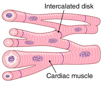
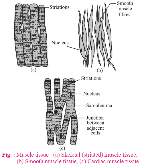
Broadly considered, human muscle—like the muscles of all vertebrates—is often divided into striated muscle, smooth muscle, and cardiac muscle.
Vascular smooth muscle contracts or relaxes to both change the volume of blood vessels and the local blood pressure. The high purity, low passage, rigorously characterized cells are performance tested prior to release. It constitutes much of the musculature of. • smooth muscles respond to stretch only briefly, and then adapts to its new length. Smooth muscle, muscle that shows no cross stripes under microscopic magnification. This is in contrast to skeletal and cardiac muscle, which have bands vascular smooth muscle helps with this second strategy. Smooth muscle tissue, unlike striated muscle, contracts slowly and automatically. Visceral muscle tissue, or smooth muscle, is tissue associated with the internal organs of the body, especially those in the abdominal cavity. Smooth muscle is a type of tissue found in the walls of hollow organs, such as the intestines, uterus and stomach. Diagrams, how muscles work in women. In this report, we assessed their roles by analyzing mice deficient of myl9, a. Vascular smooth muscle refers to the particular type of smooth muscle found within, and composing the majority of the wall of blood vessels. Human circulatory system vector illustration diagram, blood vessels scheme.
Human leg muscles diagram human leg muscle diagram anatomy body diagram. Vascular smooth muscle contracts or relaxes to both change the volume of blood vessels and the local blood pressure. Vascular smooth muscle refers to the particular type of smooth muscle found within, and composing the majority of the wall of blood vessels. Smooth muscle structure, embryonic origin, and histology. Muscular movements help the passage of materials such as blood, lymph, and food in the digestive system.
Muscular Tissue Skeletal Smooth And Cardiac Muscle Online Biology Notes from www.onlinebiologynotes.com Vascular smooth muscle refers to the particular type of smooth muscle found within, and composing the majority of the wall of blood vessels. Human mesenchymal stem cells (mscs) are used for generating smcs, and understanding the underlying regulatory mechanisms of the. Visceral muscle tissue, or smooth muscle, is tissue associated with the internal organs of the body, especially those in the abdominal cavity. Human leg muscles diagram leg muscle chart gosutalentrankco. Muscular movements help the passage of materials such as blood, lymph, and food in the digestive system. It is divided into two subgroups; • the new length however, retains its original _ seconds or minutes after it has been. Smooth muscle histology and diagram (inlet).
In this report, we assessed their roles by analyzing mice deficient of myl9, a.
Vascular smooth muscle cells (smc) line the walls of large and small arteries, arterioles & veins, and play critical we offer primary smooth muscle cells fom different tissues, species, and vascular beds. Vascular smooth muscle cell phenotypic modulation is the ability to switch phenotypic characteristics from a migratory synthetic phenotype in embryonic tissue patterning to a quiescent, contractile phenotype in maintenance of vascular tone in mature vessels. The high purity, low passage, rigorously characterized cells are performance tested prior to release. Diagrams, how muscles work in women. Learn vocabulary, terms and more with flashcards, games and other study tools. Smooth muscle is a type of tissue found in the walls of hollow organs, such as the intestines, uterus and stomach. Human circulatory system vector illustration diagram, blood vessels scheme. Human leg muscles diagram leg muscle chart gosutalentrankco. Hair transplantation procedure diagram with steps. This is in contrast to skeletal and cardiac muscle, which have bands vascular smooth muscle helps with this second strategy. Human leg muscles diagram human leg muscle diagram anatomy body diagram. It is the pen diagram of skeletal, smooth and cardiac muscle for class 10, 11 and 12. Myosin light chain kinase, smooth muscle, an enzyme which phosphorylates myosin regulatory light chains to facilitate.
This is in contrast to skeletal and cardiac muscle, which have bands vascular smooth muscle helps with this second strategy. Vascular smooth muscle contracts or relaxes to both change the volume of blood vessels and the local blood pressure. It is divided into two subgroups; Smooth muscle (also known as visceral muscle due to the locations in which they are present ) is one of the three main types of muscle tissue that exist in the human body. Vascular smooth muscle cell phenotypic modulation is the ability to switch phenotypic characteristics from a migratory synthetic phenotype in embryonic tissue patterning to a quiescent, contractile phenotype in maintenance of vascular tone in mature vessels.
Structural Organisation In Animals Biology Notes For Neet Aiims Jipmer from lh4.googleusercontent.com Get access to ncert solutions for class 11 biology chapter 20 locomotion and movement. Human leg muscles diagram leg muscle chart gosutalentrankco. Human circulatory system vector illustration diagram, blood vessels scheme. Visceral muscle tissue, or smooth muscle, is tissue associated with the internal organs of the body, especially those in the abdominal cavity. It is the pen diagram of skeletal, smooth and cardiac muscle for class 10, 11 and 12. The high purity, low passage, rigorously characterized cells are performance tested prior to release. When vascular smooth muscle relaxes, the lumen of blood vessels enlarges, allowing more. Myosin light chain kinase, smooth muscle, an enzyme which phosphorylates myosin regulatory light chains to facilitate.
Vascular smooth muscle refers to the particular type of smooth muscle found within, and composing the majority of the wall of blood vessels.
Vascular smooth muscle cell phenotypic modulation is the ability to switch phenotypic characteristics from a migratory synthetic phenotype in embryonic tissue patterning to a quiescent, contractile phenotype in maintenance of vascular tone in mature vessels. What does a smooth muscle cell structure look like? Myosin light chain kinase, smooth muscle, an enzyme which phosphorylates myosin regulatory light chains to facilitate. The key difference between multiunit and visceral smooth muscle lies in the way in which its individual cells function. Because visceral muscle is controlled by the unconscious part of the brain, it is known as involuntary muscle—it cannot be directly controlled by. Cardiac, skeletal and smooth muscles are the three types of muscles found in the human body. The trichome stain can be used to highlight smooth muscle cells (red) and background collagen (blue) in cases of spindled cell tumors. Primary human aortic smooth muscle cells (hasmc)primary human coronary artery smooth muscle cells (hcasmc)primary human pulmonary artery smooth muscle cells (hpasmc)> 500,000 cells/vial, cryopreserved at smooth muscle cell culture systems. Muscular movements help the passage of materials such as blood, lymph, and food in the digestive system. It constitutes much of the musculature of. It is divided into two subgroups; Human leg muscles diagram human leg muscle diagram anatomy body diagram. Smooth muscle tissue, unlike striated muscle, contracts slowly and automatically.
Get access to ncert solutions for class 11 biology chapter 20 locomotion and movement smooth muscle diagram. Visceral muscle tissue, or smooth muscle, is tissue associated with the internal organs of the body, especially those in the abdominal cavity.
